(Peer-Reviewed) AI-powered nonlinear optical imaging reveals protein spatial homogenization as an indicator of impaired bone quality in type 2 diabetes
Bowen Zhang 张博文 ¹, Jiangbo Pu 蒲江波 ¹, Tao Hu 胡韬 ², Junjie Zeng 曾钧杰 ³, Han Zhang 张涵 ³, Zemeng Chen 陈泽蒙 ¹, Xiang Ji 吉祥 ¹, Shuhua Yue 岳蜀华 ³, Lin Z. Li ⁴, Ting Li 李婷 ¹
¹ Biomedical Engineering Institute, Chinese Academy of Medical Sciences and Peking Union Medical College, Tianjin 300192, China
中国 天津 中国医学科学院生物医学工程研究所 北京协和医学院
² Department of Spine Surgery, Shanghai East Hospital, School of Medicine, Tongji University, Shanghai 200092, China
中国 上海 上海市东方医院(同济大学附属东方医院)脊柱外科
³ Key Laboratory of Biomechanics and Mechanobiology (Beihang University), Ministry of Education, Institute of Medical Photonics, Beijing Advanced Innovation Center for Biomedical Engineering, School of Biological Science and Medical Engineering, Beihang University, Beijing 100191, China
中国 北京 北京航空航天大学生物与医学工程学院 北京市生物医学工程高精尖创新中心 生物力学与力生物学教育部重点实验室
⁴ Britton Chance Laboratory of Redox Imaging, Department of Radiology, Perelman School of Medicine University of Pennsylvania, Philadelphia, PA 19104, USA
Opto-Electronic Advances, 2026-05-15
Abstract
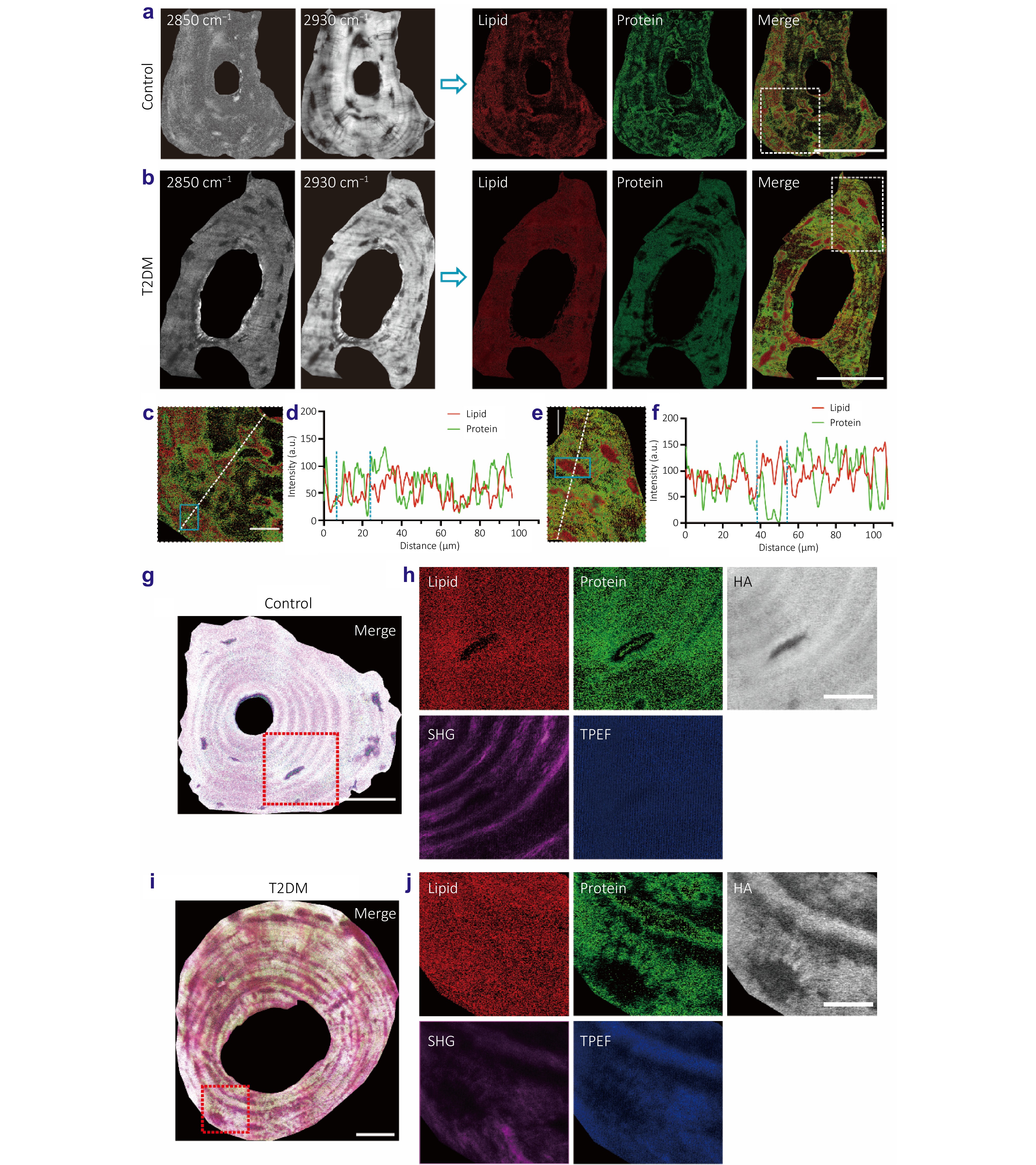
Type 2 diabetes mellitus (T2DM) significantly elevates fracture risk, a severe complication often underestimated by conventional bone mineral density (BMD) assessments. Here, we applied label-free multimodal nonlinear optical (NLO) imaging with AI-powered texture feature analysis to characterize T2DM-related bone quality alterations.
Our results identified aberrant spatial protein distribution, characterized by increased homogeneity and reduced contrast, as a distinctive pathological feature in T2DM bone. The alterations in spatial distribution were also observed in hydroxyapatite (HA) and autofluorescent metabolites. A K-nearest neighbor (KNN) model, trained on fused texture features from these three components, achieved a superior classification accuracy of 93.56% in distinguishing T2DM-related bone tissues, markedly outperforming single-component models (~70%).
This demonstrated that fused multi-component spatial distribution features offer enhanced discriminative power for quantifying T2DM-associated pathological changes. Collectively, aberrant molecular spatial distribution, particularly of protein, represents a potentially unappreciated indicator of diabetic bone quality alterations. Integrating multimodal NLO imaging with explainable AI offers a novel approach for unraveling the mechanistic underpinnings of complex pathological alterations, which not only overcomes the limitations of conventional biomarker assessment but also establishes a powerful framework for discovering new pathological targets.
Heterogeneously integrated micro-ring with SnS₂ for dual-functional optical modulation and photodetection
Jinyi Du, Lidan Lu, Xu Zhang, Bofei Zhu, Wenbo Bo, Yingjie Xu, Guang Chen, Yanlin He, Guanghui Ren, Xiaoping Lou, Zheng You, Lianqing Zhu
Opto-Electronic Advances
2026-08-25
Hardware-aware lightweight photonic spiking neural network for pattern classification
Shuiying Xiang, Yahui Zhang, Shangxuan Shi, Haowen Zhao, Dianzhuang Zheng, Xingxing Guo, Yanan Han, Ye Tian, Liyue Zhang, Yuechun Shi, Yue Hao
Opto-Electronic Advances
2026-08-25
PhyspeNet: An empirical physics-aware network for adaptive speckle reconstructive spectrometry
Junrui Liang, Min Jiang, Jun Li, Zhongming Huang, Junhong He, Yanting Guo, Yanzhao Ke, Jun Ye, Jiangming Xu, Jinyong Leng, Pu Zhou
Opto-Electronic Advances
2026-08-25
A 36 × 240 Gbps hybrid mode/wavelength division multiplexing transmitter using lithium niobate on insulator
Mingyu Zhu, Weihan Wang, Ruitao Ma, Aoyun Gao, Chun Gao, Zexu Wang, Fei Huang, Zhenyuan Bao, Dajian Liu, Jiaxuan Gan, Zejie Yu, Huan Li, Weike Zhao, Daoxin Dai
Opto-Electronic Advances
2026-08-25
Light-perception-based interactive control of an underwater digital twin hand
Jinlong Lu, Chao Zhang, Hengchang Nong, Dongying Wang, Hongyu Zhou, Junjie Weng, Yuehua Deng, Yang Yu, Qiang Bian, Jianfa Zhang, Chaofan Zhang, Zhenrong Zhang, Junbo Yang
Opto-Electronic Advances
2026-07-10