(Peer-Reviewed) Contrast-enhanced ultrasound as a valuable imaging modality for characterizing testicular lesions
Jie Yu 于洁 ¹, Xin-Hui Jiang 江鑫辉 ², Lian-Fang Du 杜联芳 ¹, Min Bai 白敏 ¹, Zhao-Jun Li 李朝军 ¹, Qiu-Sheng Shi 史秋生 ¹, Qi Jiang 蒋琪 ³, Fan Li 李凡 ¹
¹ Department of Ultrasound, Shanghai General Hospital, Shanghai Jiao Tong University School of Medicine, Shanghai 201620, China
中国 上海 上海交通大学医学院 上海市第一人民医院 超声科
² Department of Medical Ultrasound, Shanghai Baoshan Hospital of Integrated Traditional Chinese and Western Medicine, Shanghai 201999, China
中国 上海 上海市宝山区中西医结合医院 超声科
³ Department of Urology, Shanghai General Hospital, Shanghai Jiao Tong University School of Medicine, Shanghai 200080, China
中国 上海 上海交通大学医学院 上海市第一人民医院 泌尿科
Asian Journal of Andrology
, 2021-09-14
Abstract
Contrast-enhanced ultrasound (CEUS) is a new form of ultrasound (US) that can dynamically display microvessels in a highly sensitive manner. The purpose of this study was to investigate the efficacy of CEUS for characterizing testicular lesions in comparison with conventional US. Forty-seven patients with testicular lesions were enrolled.
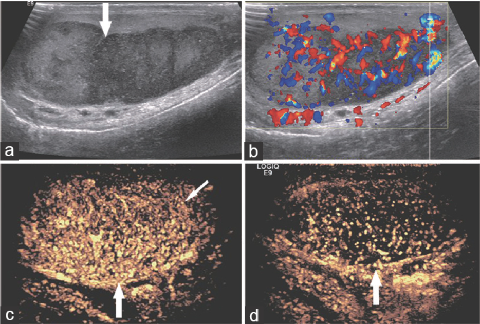
The histopathology results revealed that 31 cases were neoplastic (11 cases of seminomas, 8 nonseminomatous germ cell tumors, 8 lymphomas, 2 Leydig cell tumors, and 2 nonspecific tumors), and 16 cases were nonneoplastic (8 cases of infarctions, 3 epidermoid cysts, and 5 inflammation). The indicators of shallow lobulated morphology and cystic-solid echogenicity on conventional US were suggestive of germ cell tumors. More indicators on CEUS were found to be useful for characterizing testicular lesions. All the neoplastic lesions showed hyperenhancement on CEUS. Moreover, germ cell tumors presented with heterogeneous enhancement (73.7%, 14/19), a twisted blood vessel pattern, rapid wash-in and wash-out, and peripheral rim hyperenhancement signs. Lymphoma was characterized by nonbranching linear vessel patterns (87.5%, 7/8), rapid wash-in and slow wash-out.
In nonneoplastic lesions, infarction and epidermoid cysts showed no enhancement, and abscesses were observed with marginal irregular enhancement. The sensitivity, specificity, and accuracy of CEUS for differentiating between neoplastic and nonneoplastic lesions were 100%, 93.8%, and 97.9%, respectively, and these values were higher than those for conventional US (90.3%, 62.5%, and 80.9%, respectively). CEUS can sensitively reflect the microvascular perfusion in testicular lesions and offers high accuracy for characterizing them.
Flicker minimization in power-saving displays enabled by measurement of difference in flexoelectric coefficients and displacement-current in positive dielectric anisotropy liquid crystals
Junho Jung, HaYoung Jung, GyuRi Choi, HanByeol Park, Sun-Mi Park, Ki-Sun Kwon, Heui-Seok Jin, Dong-Jin Lee, Hoon Jeong, JeongKi Park, Byeong Koo Kim, Seung Hee Lee, MinSu Kim
Opto-Electronic Advances
2025-09-25
Dual-frequency angular-multiplexed fringe projection profilometry with deep learning: breaking hardware limits for ultra-high-speed 3D imaging
Wenwu Chen, Yifan Liu, Shijie Feng, Wei Yin, Jiaming Qian, Yixuan Li, Hang Zhang, Maciej Trusiak, Malgorzata Kujawinska, Qian Chen, Chao Zuo
Opto-Electronic Advances
2025-09-25


