(Peer-Reviewed) Experimental investigation of early assessment of corpora cavernosa fibrosis with two-dimensional shear wave elastography
Li Yu ¹, Wan-Ting Rao ², Jing-Dong Tang 汤敬东 ³, Jin-Fang Xing 邢晋放 ² ³
¹ Department of Special Inspection, Shanghai Shibei Hospital of Jing'an District, Shanghai 200435, China
中国 上海 上海市静安区市北医院特检科
² Department of Medical Ultrasound, Fudan University Pudong Medical Center, Shanghai 201399, China
中国 上海 复旦大学附属浦东医院超声医学科
³ Shanghai Key Laboratory of Vascular Lesions Regulation and Remodeling, Fudan University Pudong Medical Center, Shanghai 201399, China
中国 上海 复旦大学附属浦东医院 上海市血管病变调控与重塑重点实验室
Asian Journal of Andrology, 2021-09-03
Abstract
This study explored the usefulness of two-dimensional shear wave elastography (2D-SWE) in the early assessment of corpora cavernosa fibrosis (CCF). New Zealand male rabbits were randomly assigned to an experimental group or a control group.
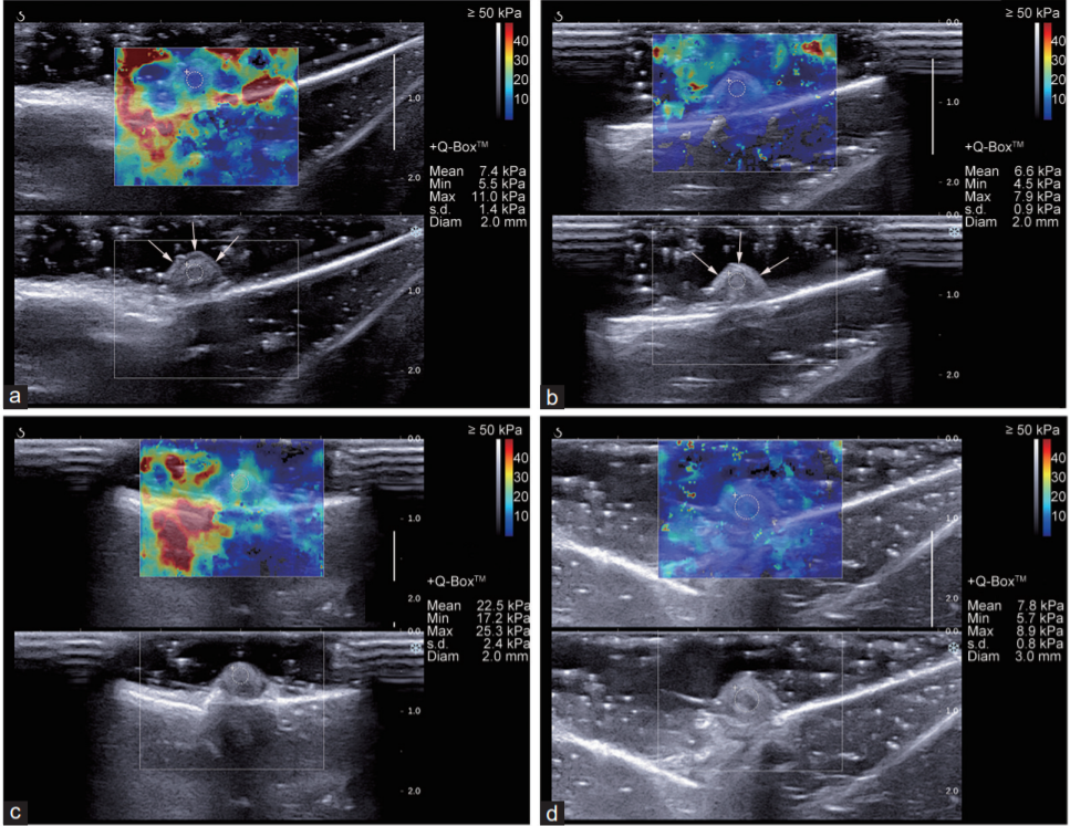
Recombinant human transforming growth factor beta 1 (TGF-β1) was injected into the dorsal penis tissue of rabbits in the experimental group. Conventional ultrasound and 2D-SWE examinations were performed before and 20 days after injection. Penile histological analysis was performed by hematoxylin–eosin staining, sirius red staining, and immunohistochemistry. Measurement of 2D-SWE examination results was performed using shear wave elastography quantitative measurement (SWQ). Histological analysis outcomes were the proportion of smooth muscle cells (SMCs), collagen fibers (CFs), collagen type I (Col I), and collagen type III (Col III), as well as the SMCs/CFs ratio, measured by sirius red staining.
Other histological analysis outcomes were the positive area proportion (PAP) of TGF-β1 (PAPT), fibronectin (PAPF), and Col III (PAPC), measured by immunohistochemistry. After recombinant human TGF-β1 injection, SWQ was higher in the experimental group than that in the control group (P < 0.001); however, there were no differences in conventional ultrasound results. There were significant differences in histological outcomes between the two groups (all P < 0.05). These results indicated that 2D-SWE was superior for identifying early histological changes in CCF.
A review on optical torques: from engineered light fields to objects
Tao He, Jingyao Zhang, Din Ping Tsai, Junxiao Zhou, Haiyang Huang, Weicheng Yi, Zeyong Wei Yan Zu, Qinghua Song, Zhanshan Wang, Cheng-Wei Qiu, Yuzhi Shi, Xinbin Cheng
Opto-Electronic Science
2025-11-25
Flicker minimization in power-saving displays enabled by measurement of difference in flexoelectric coefficients and displacement-current in positive dielectric anisotropy liquid crystals
Junho Jung, HaYoung Jung, GyuRi Choi, HanByeol Park, Sun-Mi Park, Ki-Sun Kwon, Heui-Seok Jin, Dong-Jin Lee, Hoon Jeong, JeongKi Park, Byeong Koo Kim, Seung Hee Lee, MinSu Kim
Opto-Electronic Advances
2025-09-25


